
LNP Protein Corona: How Blood Proteins Reshape RNA Delivery

The LNP protein corona raises a practical question for RNA delivery: after a carefully formulated particle enters blood, what surface does a cell actually encounter?
Lipid chemistry and targeting ligands matter. So do the proteins that associate with the particle in biological fluids. Even after cellular entry, RNA must cross an endosomal membrane. Understanding that journey requires connecting the protein corona, apparent LNP pKa, and endosomal escape while measuring each step separately.

Why RNA needs a delivery system in the first place
mRNA supplies instructions for protein production; siRNA can reduce a specific RNA through the cell's RNA-interference machinery. Both face physical barriers: RNA is relatively large, hydrophilic, and negatively charged, crosses membranes inefficiently on its own, and can undergo extracellular nuclease degradation.
Activity after experimental introduction into a cell does not establish activity following direct injection. A formulation must preserve sufficient intact cargo, reach the intended cells, and deliver it to the appropriate intracellular location. LNPs are one approach, rather than a requirement for every RNA medicine. Delivery background
Three questions help organize the problem: which organ should encounter the RNA, which cells should use it, and what should those cells do? The spleen contains T cells, B cells, macrophages, and dendritic cells, among others. Strong splenic signal does not identify which population received functional RNA.
What the four common LNP components contribute
Many RNA-LNPs combine four lipid classes. Their roles overlap, and the table is a starting framework rather than a mandatory recipe for every particle. Lipid-design background
Component | Examples | Design role | Connection to this article |
Ionizable lipid | DLin-MC3-DMA, SM-102, ALC-0315 | pH-dependent protonation, RNA encapsulation, and intracellular membrane interactions | A major variable in pKa, structure, and escape |
Helper phospholipid | DSPC, DOPE | Assembly, organization, and membrane packing | Changing it can alter overall performance |
Cholesterol or another sterol | Cholesterol | Lipid packing, fluidity, and structural stability | Changes the environment for interfacial and membrane interactions |
PEG-lipid | Lipid-anchored PEG | Assembly, colloidal stability, and surface shielding | Configuration and desorption influence protein and cell contact |
Two particles containing SM-102 need not have identical targeting. Other components, ratios, manufacturing, and cargo can differ. The particle is an interacting molecular assembly rather than a fixed container whose behavior is specified by one ingredient.
Why the LNP protein corona matters for targeting
The term protein corona describes proteins associated with a nanoparticle following exposure to biological fluids. The interface depends on particle composition, surface properties, PEG configuration, and the surrounding environment.
Li and colleagues' 2026 review places corona biology alongside lipid architecture, formulation, ligands, and administration route. This is a useful engineering perspective: organ preference emerges from interacting variables. A formulation's behavior still needs validation across species, routes, and disease settings. Review

ApoE provides an example of endogenous recognition
Akinc and colleagues used recombinant protein and ApoE- and LDLR-deficient systems to support an endogenous targeting role for ApoE in the ionizable LNPs they tested. Dependence differed across carrier types. Original study
That distinction matters. A particle without an attached antibody may still engage biological recognition through endogenous proteins. It does not follow that every LNP uses the same pathway.
A clinical example: why patisiran is intended for hepatocytes
Hepatic delivery can be the desired outcome. Patisiran, formulated as the Onpattro lipid complex, delivers siRNA to hepatocytes to reduce TTR mRNA and subsequent TTR protein through RNA interference. Product description and mechanism
This separates the cell receiving RNA from the tissues affected by the disease-associated protein. RNA need not reach every affected tissue to alter a relevant protein source. Changing the intended recipient to a T cell, pulmonary cell, or brain cell requires a different delivery and validation strategy.
SORT connects formulation chemistry with protein interactions
A 2021 SORT study connected lipid composition, biodistribution, apparent pKa, and serum interactions in a proposed endogenous targeting mechanism, while retaining the possibility of additional mechanisms. Mechanistic study
A subsequent lung-delivery study found that SORT lipid structure was associated with changes in both corona composition and organ selectivity. No single protein independently explained lung selectivity across the formulations. 2023 study
Proteomics can identify mechanistic candidates. It cannot, by itself, establish that the most abundant associated protein causes targeting. Functional perturbation is needed to distinguish a marker of a successful formulation from a contributor to its activity.
A preclinical example: redirecting a platform toward lung or spleen
The original 2020 SORT study added supplementary lipids to existing LNP platforms. Tested permanently cationic components, including DOTAP, biased expression toward lung; anionic components, including 18PA, biased it toward spleen. Delivery was assessed in mice through reporters, cell analysis, and gene-editing experiments. Original SORT study
DOTAP is not a lung-recognizing antibody. The example demonstrates formulation-level redirection of functional delivery under the tested conditions; it does not establish that adding the same ingredient to any LNP will reproduce that selectivity in humans.
What does abundance in a corona dataset actually mean?
The denominator matters. A protein's share of the recovered mass-spectrometry signal differs from its enrichment relative to the starting plasma. Neither measurement alone establishes receptor engagement, orientation, conformation, or accessibility.
Sample handling matters too. Serum and plasma are different starting materials. Incubation, separation, and washing influence the recovered profile. The terms hard and soft corona operationally distinguish more persistent association from more exchangeable interactions; they should not be interpreted as two permanent anatomical layers. Li and colleagues highlight characterization limits that complicate mechanistic interpretation. Review
I would look for fluid-background and particle-free controls, recovery checks, and confirmation of particle integrity before interpreting a proteomic profile as a functional interface.
Apparent LNP pKa: which ionization behavior is being measured?
For a simplified basic site, B + H⁺ ⇌ BH⁺, pKa refers to dissociation of the conjugate acid BH⁺. The intrinsic pKa of a free molecule or model compound cannot simply be substituted for the apparent pKa of an assembled LNP.
Local hydration, neighboring lipids, organization, and electrostatics influence protonation. A 2026 constant-pH molecular dynamics study illustrates these dependencies in simplified membrane systems, providing a molecular interpretation rather than a direct measurement of a complete LNP in humans. Model-membrane study
TNS fluorescence titration indirectly probes positive charge at the particle interface. Its fitted midpoint is an operational apparent pKa, not a count demonstrating that half of all amines throughout the particle are protonated. TNS formulation study
Zeta potential describes electrokinetic potential near the slipping plane in a specified medium. A single measurement at pH 7.4 cannot replace a titration curve. Comparisons should report buffer, ionic strength, temperature, probe and particle concentrations, incubation, and fitting. Multiple titratable components or non-sigmoidal curves also complicate assigning one number to one chemical group. The pH at which zeta potential crosses zero is an operational isoelectric point, not necessarily the apparent pKa. Ionization study
A simplified calculation explains the acidification response
Consider three environments first. Acidic formulation conditions help some ionizable lipids associate with RNA. Near physiological pH, many such formulations seek reduced exposed positive charge. After uptake, endosomal acidification can promote renewed membrane interactions. This is a design strategy, not a claim that every site switches synchronously or that circulating particles have no charge or protein interactions. Formulation background
For one class of independent protonatable sites, the Henderson–Hasselbalch relationship gives:
Protonated fraction f = 1 / [1 + 10^(pH − pKa)].
Using an assumed pKa of 6.4 yields the following rounded values:
Assumed environmental pH | Idealized protonated-site fraction |
7.4 | About 9% |
6.5 | About 44% |
5.5 | About 89% |
This is an educational calculation, not measured LNP data or a prediction of surface charge or escape. Applying an apparent pKa to this idealized equation does not resolve every lipid's protonation within a heterogeneous particle.
Why 6.2–6.5 is not a universal optimum
Jayaraman and colleagues identified an approximately 6.2–6.5 optimum in their intravenous hepatic siRNA-silencing system; lipids with similar pKa still differed in potency. Hassett and colleagues found route-dependent behavior and a different preferred range in their tested intramuscular mRNA vaccine formulations. Hepatic siRNA study, intramuscular mRNA study
An optimum must be attached to its payload, route, cell, and endpoint. SORT formulations containing permanent cationic or anionic components also cannot be reduced to one amine's titration. Apparent pKa is a useful descriptor, not an organ address or a guarantee of escape.
Cargo deserves attention as well. A 2026 study found payload-size- and structure-dependent apparent pKa changes in tested formulations. Switching a reporter RNA to a therapeutic cargo therefore warrants characterization rather than assuming unchanged ionization. Original study
Endosomal escape: cellular entry is still short of functional delivery
Following endocytosis, RNA generally remains separated from the cytosol. mRNA needs cytosolic access for translation; siRNA needs access to the RNA-interference machinery. Uptake, cytosolic RNA, and downstream activity are distinct measurements.
Endosomes are normal cellular sorting and transport compartments. Internalized material can be routed toward recycling, further transport, or degradation. For RNA delivery, the obstacle is the membrane separating the endosomal lumen from the cytosol. Numerous intracellular fluorescent puncta may represent cargo trapped in those compartments rather than abundant cytosolic RNA. Trafficking and imaging study
A commonly investigated model connects endosomal acidification with lipid protonation, interaction with accessible anionic membrane lipids, and changes in packing and curvature. Non-bilayer arrangements, including inverted hexagonal HII phases, are supported in some systems. A 2025 study combining lipid comparisons, simulations, and ³¹P NMR illustrates why spatial conformation can distinguish cytosolic delivery even among lipids with similar pKa. Original study
These are formulation- and membrane-dependent processes. A model membrane's phase transition does not alone demonstrate intact RNA release in cells. Nor should proton-sponge rupture be presented as an established universal LNP mechanism.
Gilleron and colleagues estimated approximately 1–2% cytosolic escape of internalized siRNA in their tested LNP systems, within a limited trafficking window. This influential result illustrates a bottleneck, not a fixed efficiency for all modern LNPs, mRNAs, or target cells. 2013 study
Wittrup and colleagues directly imaged siRNA release and linked it with subsequent knockdown in a lipoplex system. For the smaller LNPs, they instead used Galectin-8 recruitment and changes in cargo fluorescence as indirect readouts of release. The results implicate maturing endosomes while emphasizing the distinction between direct observation and surrogate measurements, and the importance of where and when release occurs. 2015 study

Targeting moieties and formulation must support productive delivery together
Antibodies, antibody fragments, peptides, and other targeting moieties can increase an LNP's opportunity to engage a desired cell or receptor. Receptor binding addresses only part of delivery: internalization, intracellular routing, and release of intact mRNA from endosomes still determine whether that engagement becomes useful expression.
A targeting moiety can support recognition and uptake; the formulation must also support RNA protection, appropriate trafficking, endosomal escape, and unpacking. Productive delivery requires these steps to work together. The CD5-directed example discussed later evaluates delivery and cellular expression experimentally; attaching a CD5 antibody does not establish that any underlying formulation can generate CAR T cells efficiently. Original cell-targeting study
A good formulation therefore means more than favorable particle size, encapsulation, or uptake. Relevant properties include ionizable-lipid protonation and membrane interactions, helper-lipid and sterol organization, PEG shielding and desorption, and RNA integrity and release. Apparent pKa informs characterization but cannot guarantee escape. Performance should be judged through functional delivery and acceptable injury in the intended cells. Formulation evaluation linking uptake, escape, and translation
Consider a hypothetical screen in which adding a ligand increases target-cell uptake without increasing protein expression. After excluding surface adhesion and labeling artifacts, this supports increased uptake. Improved receptor-specific recognition still requires binding or receptor-perturbation experiments, and productive mRNA delivery has not yet been shown to improve. Trafficking, cytosolic release of intact RNA, and translation warrant investigation before simply increasing ligand density further.

Co-optimize targeting and escape while distinguishing their contributions
These are not entirely independent modules. Receptor engagement can alter intracellular routing, while conjugation, ligand density, and PEG configuration may change the interface and protein interactions. The final surface-modified formulation therefore needs its own performance assessment. I would consider the following comparison, rather than assume a specific product has completed it:
Comparison group | Main question |
Original formulation without targeting moiety | What are baseline uptake, cytosolic RNA, and expression? |
Same formulation with targeting moiety | Do recognition and function improve together at comparable particle quality? |
Optimized formulation without targeting moiety | How much improvement comes from formulation changes alone? |
Optimized formulation with targeting moiety | Can selectivity, function, and tolerability be maintained together? |
Interpretation requires attention to RNA dose, particle quality, receptor expression, non-target cells, and viability, with nonbinding-ligand or receptor-blockade controls where appropriate. A claim of better escape per unit of internalized RNA additionally requires quantifying uptake and cytosolic release; total protein signal alone cannot establish it.
The development objective is sufficient intact mRNA performing its intended function in the desired cells. A targeting moiety still needs a formulation that supports endosomal escape, and a potent formulation still needs evidence of adequate target-cell selectivity and function. This is an integrated development interpretation, not an outcome guaranteed by one ligand or pKa value.
Connecting corona, pKa, and escape without conflating them
I would track fluid interactions → cell engagement and uptake → endosomal trafficking → RNA release and unpacking → cytosolic function.
Corona composition may influence cell and receptor interactions, potentially changing uptake and trafficking. Whether it improves escape requires direct testing. Apparent pKa informs protonation behavior; lipid conformation, membrane contact, PEG shielding and desorption, and RNA unpacking also matter. Greater uptake need not mean more usable cytosolic RNA.
For example, similar uptake but different protein expression across serum conditions should prompt investigation of trafficking, escape, RNA integrity, and translation. It does not, by itself, establish that the corona improved escape. This is an experimental reasoning framework, not a universally validated pathway.
A hypothetical example: more uptake can yield less cytosolic RNA
The following numbers are entirely illustrative. Assume intact RNA, exclusion of surface-bound material, and a definable fraction of internalized RNA reaching the cytosol:
Assumed measurement per cell | Formulation A | Formulation B |
Internalized RNA molecules | 10,000 | 5,000 |
Fraction reaching the cytosol | 1% | 5% |
Calculated cytosolic RNA molecules | 100 | 250 |
These are neither study results nor performance targets. A has twice the uptake but fewer cytosolic molecules because of its lower release fraction. Even this arithmetic does not predict protein output or efficacy: unpacking, RNA integrity, translation, and cell state remain relevant.
Selecting solely on cell-associated fluorescence could therefore favor particles that accumulate in endosomes over particles that provide more functional delivery.
Which measurements support an escape claim?
Measurement | Main question | What it cannot establish alone |
Labeled RNA/lipid flow cytometry or imaging | Cell-associated signal and uptake | Surface adhesion must be excluded; lipid signal is not intact RNA |
Dynamic Rab5, Rab7, or LAMP1 colocalization | Intracellular compartment and trafficking | Reduced colocalization is not a quantitative escape measurement |
Galectin-8/9 recruitment | Endosomal membrane damage | Number of intact released RNA molecules or translation competence |
Cytosolic fractionation and RNA quantification | RNA detected in the cytosolic fraction | Endosomal contamination and RNA integrity need controls |
Protein expression, knockdown, or editing | Functional delivery | The bottleneck's location, independently of RNA design and cell state |
Munson and colleagues combined uptake, Galectin-9 recruitment, and mRNA translation in a screening platform and observed cell-line differences. Complementary measurements are more informative than a single fluorescent image. Original assay study
Time- and dose-matched comparisons, negative controls, viability, and membrane-injury measurements also matter. Increasing damage is not automatically an improvement in delivery.
Does corona always mean an outer protein shell?
A 2026 Nano Letters study complicates a familiar illustration. In the tested LNP systems, imaging did not reveal a conventional peripheral protein shell; experiments and simulations supported lipoprotein integration and membrane remodeling. The authors also noted that some electron-dense structures could represent membrane blebs, or blebs together with fused lipoproteins; the images alone do not establish the identity of every structure. Original study
This does not settle the architecture of all LNP–protein interfaces. It separates two questions: which proteins accompany a particle, and how those proteins participate structurally. The discussion here is limited to the accessible abstract and figure descriptions, rather than the complete supplementary experiments.
Connecting this with ERTLNP and CAR-NK
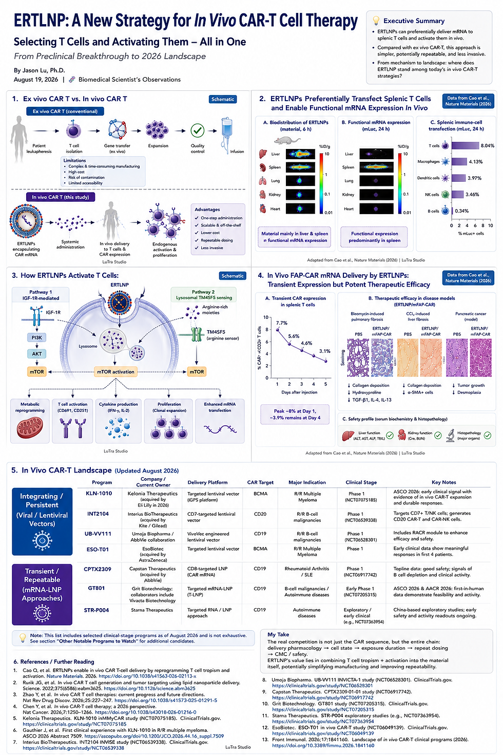
Our earlier ERTLNP article discussed material design and T-cell state; the CAR-NK topic explored receptor-directed delivery and immune regulation.
Corona biology adds the interface that develops in biological fluids. These perspectives can coexist, but mechanisms cannot be transferred between formulations without direct evidence. A protein implicated in SORT does not automatically explain ERTLNP behavior.
A cell-targeting example: CD5 and FAP have different jobs
Rurik and colleagues' 2022 study used CD5-targeted LNPs to deliver FAP CAR mRNA to T cells and evaluated the approach in a mouse cardiac-injury model. CD5 served the delivery side of recognition; FAP was the target recognized by the resulting CAR. Original study
Distinguishing these layers prevents confusion between the cell receiving RNA and the cell subsequently attacked by an engineered immune cell. This example clarifies targeting terminology; it does not establish a corona mechanism or human cardiac efficacy.
What would make a targeting mechanism convincing?
I would look for an experimental chain connecting the material, candidate protein, receptor, and functional outcome. The appropriate experiments depend on the system.
Question | Useful evidence | Interpretation limit |
Which proteins accompany the recovered particles? | Proteomics with appropriate separation and background controls | Association does not establish function |
Does the candidate contribute to delivery? | Depletion, blockade, or genetic perturbation | The intervention may also change the fluid or particle |
Can activity be restored? | Protein add-back with controls | Concentration, species, and protein state matter |
Is a receptor involved? | Receptor perturbation and cell-resolved analysis | Uptake must be distinguished from functional RNA activity |
Organ-level reporter expression helps screen formulations. A mechanism also needs evidence about the cells involved and whether changing the proposed interaction changes the intended function.
Why the biological environment needs its own validation
A 2025 study using liver-humanized mice and primary human hepatocytes demonstrated serum-related species restrictions on LNP delivery. This is a warning about model dependence, not proof that a lipid cannot work in humans or that results transfer to intramuscular vaccines. Original study
The development implication is to examine how performance depends on the fluid and cellular environment. Relevant comparisons may include different biological fluid sources, disease conditions, and repeat-dose settings, together with immune responses and off-target exposure.
A CMC question about reproducible biological performance
If a formulation depends on protein interactions, I would ask whether batches with similar size and encapsulation also maintain similar functional delivery to the intended cells. Storage and manufacturing changes should be assessed against the product's meaningful biological activity.
Apparent pKa can contribute to characterization when the method is reproducible and sensitive to relevant changes. An escape assay must specify whether it measures membrane injury, cytosolic RNA, or downstream function. Matching pKa does not replace cell-level potency comparisons; matching reporter expression can also conceal different uptake or toxicity.
This is a development inference, not a claim that every LNP requires corona proteomics as a release assay. Analytical strategies should fit the particular product and mechanism and demonstrate sensitivity to consequential changes.
The biological environment continues to shape a particle after manufacturing. Adding pKa and endosomal escape completes the development question: how cells recognize the particle, where its charge and membrane interactions change, and how much intact RNA reaches the cytosol to perform the intended function.
References
1. Li J, et al. Emerging Lipid Nanoparticle Targeting Strategies for Extrahepatic Delivery of RNA Therapeutics. Advanced Materials. 2026. DOI
2. Akinc A, et al. Targeted Delivery of RNAi Therapeutics With Endogenous and Exogenous Ligand-Based Mechanisms. Molecular Therapy. 2010. DOI
3. Dilliard SA, Cheng Q, Siegwart DJ. On the mechanism of tissue-specific mRNA delivery by selective organ targeting nanoparticles. PNAS. 2021. DOI
4. Dilliard SA, et al. The interplay of quaternary ammonium lipid structure and protein corona on lung-specific mRNA delivery by selective organ targeting (SORT) nanoparticles. Journal of Controlled Release. 2023. DOI
5. Lipid Nanoparticle Protein Coronas Arise through Lipoprotein Fusion Rather Than Shell-like Adsorption. Nano Letters. 2026. DOI
6. Stone D, et al. Serum factors create species-specific barriers to hepatic gene transfer by lipid nanoparticles in liver-humanized mice. Molecular Therapy Methods & Clinical Development. 2025. DOI
7. Jayaraman M, et al. Maximizing the Potency of siRNA Lipid Nanoparticles for Hepatic Gene Silencing In Vivo. Angewandte Chemie International Edition. 2012. DOI
8. Hassett KJ, et al. Optimization of Lipid Nanoparticles for Intramuscular Administration of mRNA Vaccines. Molecular Therapy Nucleic Acids. 2019. DOI
9. Trollmann MFW, Rossetti P, Böckmann RA. Membrane environment sets the functional pKa of ionizable lipids. Biophysical Journal. 2026. DOI
10. Gilleron J, et al. Image-based analysis of lipid nanoparticle-mediated siRNA delivery, intracellular trafficking and endosomal escape. Nature Biotechnology. 2013. DOI
11. Wittrup A, et al. Visualizing lipid-formulated siRNA release from endosomes and target gene knockdown. Nature Biotechnology. 2015. DOI
12. Munson MJ, et al. A high-throughput Galectin-9 imaging assay for quantifying nanoparticle uptake, endosomal escape and functional RNA delivery. Communications Biology. 2021. DOI
13. Spatial Conformation of Ionizable Lipids Regulates Endosomal Membrane Disruption. Journal of the American Chemical Society. 2025. DOI
14. Carrasco MJ, et al. Ionization and structural properties of mRNA lipid nanoparticles influence expression in intramuscular and intravascular administration. Communications Biology. 2021. DOI
15. Priss A, et al. RNA Size and Structure Modulate the Apparent pKa of Ionizable Lipid Nanoparticles. Bioconjugate Chemistry. 2026. DOI
16. ONPATTRO (patisiran) lipid complex injection: Prescribing Information. Sections 11 and 12.1. DailyMed
17. Cheng Q, et al. Selective organ targeting (SORT) nanoparticles for tissue-specific mRNA delivery and CRISPR-Cas gene editing. Nature Nanotechnology. 2020. DOI
18. Rurik JG, et al. CAR T cells produced in vivo to treat cardiac injury. Science. 2022. DOI
Abbreviations
LNP, lipid nanoparticle; mRNA, messenger RNA; siRNA, small interfering RNA; ApoE, apolipoprotein E; LDLR, low-density lipoprotein receptor; PEG, polyethylene glycol; SORT, selective organ targeting; CMC, chemistry, manufacturing, and controls; pKa, negative logarithm of the acid dissociation constant, with apparent LNP pKa dependent on formulation and measurement; TNS, fluorescent probe of interfacial positive charge; NMR, nuclear magnetic resonance; HII phase, inverted hexagonal non-bilayer arrangement; Rab5/Rab7, endosomal trafficking proteins; LAMP1, membrane protein commonly used to identify late endosomal/lysosomal compartments; galectins, carbohydrate-binding proteins, some of which report endosomal membrane injury; TTR, transthyretin; CAR, chimeric antigen receptor; FAP, fibroblast activation protein; DSPC/DOPE, common helper phospholipids; DOTAP/18PA, permanently cationic/anionic lipids used in the SORT example.
Cited evidence last rechecked November 4, 2026, America/New_York. This recheck covers the article's existing claims and citations, rather than a comprehensive literature update. General scientific and development information; not individual medical or product-specific regulatory advice.


Comments